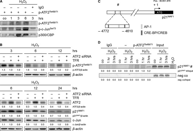
Fig. 4.
ATF2 regulates the expression of p21WAF1 and c-Jun, and p-ATF2Thr69/71 directly binds to the p21WAF1 promoter in H2O2-treated TE7 cells (250 μM). (A) p-ATF2Thr69/71 interacts with p-c-JunSer73 to form the AP-1 complex. In addition, p300 and CBP were found as p-ATF2Thr69/71 interaction partners. Cells subjected to H2O2 were lysed, and p-ATF2Thr69/71 was immunoprecipitated using anti-p-ATF2Thr69/71 antibody. Rabbit IgG was used as negative control. Precipitated lysates were immunoblotted for p-ATF2Thr69/71, p-c-JunSer73 and p300/CBP. (B) ATF2 knockdown causes a reduction in p-ATF2Thr69/71, ATF2, p21WAF1 and c-Jun protein expression. Cells were transfected with ATF2 siRNA and transfection reagent (TFR) for 7 hrs prior to H2O2 treatment. Thereafter, cells were grown for 3, 6, 12 and 24 hrs. Lysates were immunoblotted for p-ATF2Thr69/71, ATF2, p21WAF1 and c-Jun. β-actin was used as loading control. Fold expression changes are given below the blots. (C) Schematic illustration of the p21WAF1 promoter shows the putative ATF2-binding site (#), including the binding sequences AP-1, and CRE-BP/CREB −4772 to −4610 bp relative to the transcription start (+ 1; ATG, position 36.651.879), which was used for amplification. (D) p-ATF2Thr69/71 binds to the −4772 to −4610 bp p21WAF1 promoter region. Cells were treated with H2O2 and grown for 1 and 3 hrs. Cells were lysed, and p-ATF2Thr69/71 was immunoprecipitated using anti-p-ATF2Thr69/71 antibody. The target region (#) in the p21WAF1 promoter was analysed by semi-quantitative PCR. Rabbit IgG instead of anti-p-ATF2Thr69/71, an additional sample without antibody, and H2O served as negative controls. Fold PCR product accumulation is given below the gel photos.

