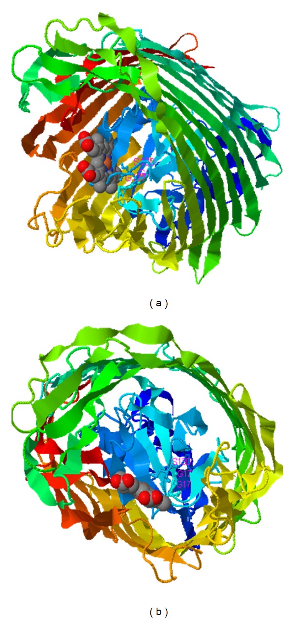
Figure 6.

BauA ligand binding sites predicted by COFACTOR. (a) and (b) show BauA structure in contact with siderophore ligand from lateral and top views, respectively. BauA is shown in ribbon and ligand in the space filling model. Conserved residues especially R(67), W(68), and F(93) from cork domain and G(303), L(305), and D(344) from barrel are involved in iron binding site.
