
Figure 1.
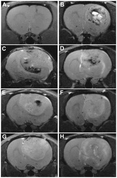
Representative T2-weighted images for gliomas from astrocyte-injected controls (Ctrl) (at 29 days following cell injection) (A), ENU (at 43 weeks of age) (B), GL261 (at 23 days following cell injection) (C), RG2 (at 17 days following cell injection) (D), U87 (at 16 days following cell injection) (E), 9L/lacZ (at 37 days following cell injection) (F), C6 (at 17 days following cell injection) (G) and F98 (at 20 days following cell injection) (H) models.