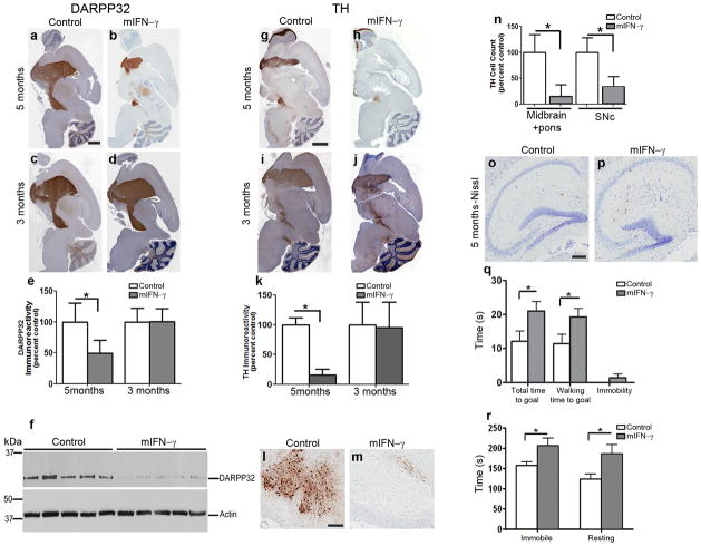
Fig 2. Selective and age-progressive nigrostriatal degeneration in rAAV2/1-mIFN-γ expressing wild type mice.
a–e. DARPP32 immunostained brain sections of 5 month old mice (a–b) or 3 month old mice (c–d) expressing rAAV2/1-mIFN-γ (b, d) or rAAV2/1-EGFP (Control) (a, c). Densitometric analysis of immunostaining is shown (e). Scale Bar, 600μm. (n=5/group; *p<0.05). Data represents mean + s.d.
f. Immunoblot showing DARPP32 and β-Actin (loading control) in 5 month old rAAV2/1-mIFN-γ expressing mice and control mice. The full length blot is depicted in Supplementary Figure 5. (n=5/group; *p<0.001).
g–k. TH immunostained brain sections of 5 month old mice (g–h) and 3 month old mice (i–j) expressing rAAV2/1-mIFN-γ (h, j) or EGFP (Control) (g, i). Densitometric analysis of immunoreactivity is shown (k). Scale Bar, 600μm. (n=5/group; *p<0.001). Data represents mean + s.d.
l–n. TH immunostained neurons in the SNc of 5 month old mIFN-γ (m) and EGFP (l) mice. Counts of TH immunoreactive cell bodies in SNc (n, “SNc”, n=5/group) and entire midbrain is depicted (n, “Midbrain+pons”; n=3/group). Scale bar, 120μm. (*p<0.001). Data represents mean + s.d.
o–p. Representative Nissl stained hippocampus from 5 month old mIFN-γ (p) and EGFP (o) mice. Scale Bar, 125μm. (n=5/group).
q–r. Locomotor and behavioral impairment is evident in 8 month old mIFN-γ mice compared to controls in the beam crossing test (q) and open field test (r). (n=8–12/group; *p<0.05). Data represents mean + s.e.m.

