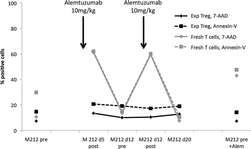
Figure 4. Alemtuzumab-containing serum, taken immediately after mAb infusion, does not kill expanded Treg.
Monkey serum was drawn pre-alemtuzumab, immediately after a second dose (10mg/kg), immediately before and after a third dose (10mg/kg), and 1 week after the third dose. These sera were incubated with freshly-isolated monkey T cells and expanded autologous Treg. The percentages of Annexin-V and 7-AAD positive cells are shown. Arrows show time-points when alemtuzumab was infused. Fresh T cells showed an increase in apoptosis and cell killing in response to high concentrations of alemtuzumab in the blood (d5 post-alemtuzumab, d12 post-alemtuzumab), as well as when exposed to pre-alemtuzumab serum to which 10 µg/ml of alemtuzumab had been added (far right). Values returned to baseline levels of apoptosis and killing when fresh T cells were incubated with serum obtained 1 week after alemtuzumab infusion (d12, d20), indicating that serum contained high concentrations of alemtuzumab early after infusion. Expanded autologous Treg showed no increase in apoptosis or killing when exposed to a high concentration of alemtuzumab in serum, whether the serum was drawn from an alemtuzumab-treated monkey, or whether alemtuzumab had been added to the serum in vitro (far right).

