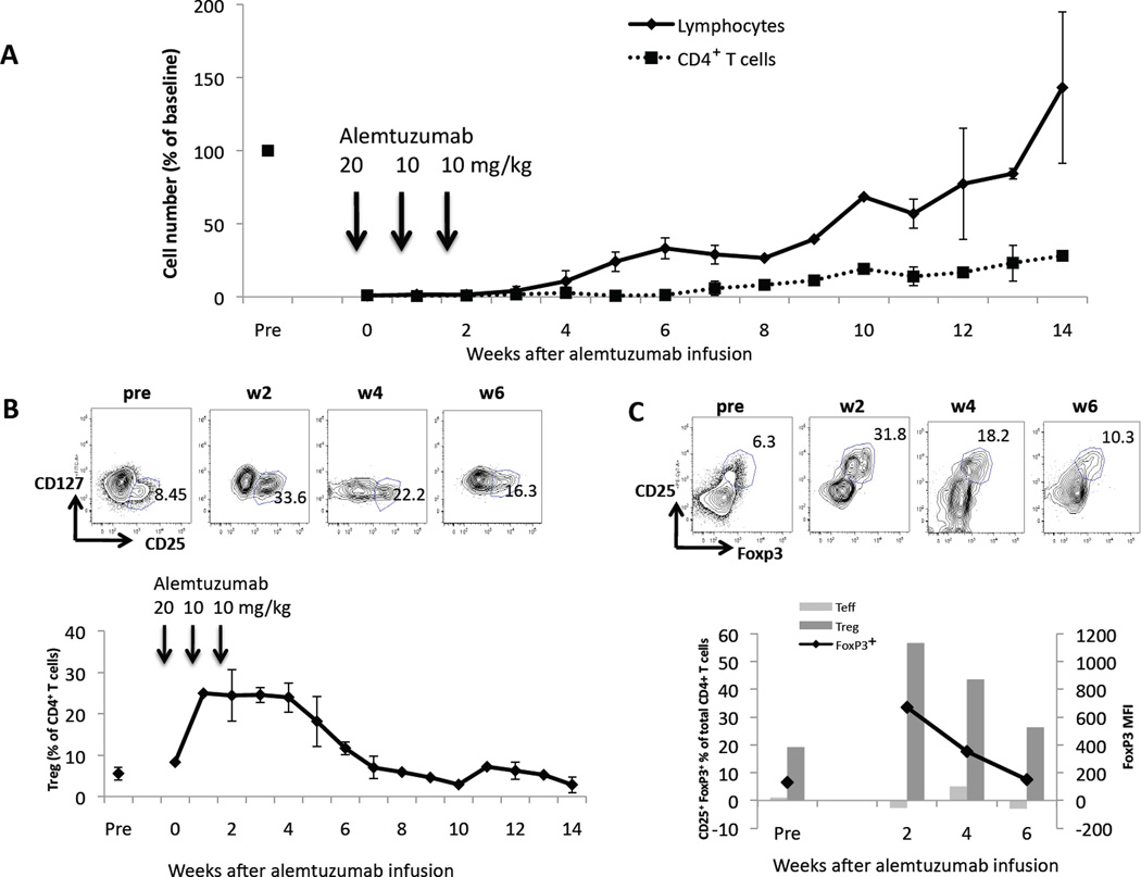
Figure 5. Relative increase in circulating Treg after infusion of alemtuzumab.
Monkeys that had received a heart transplant on d 0 and 3 doses of alemtuzumab on d -2, 5, and 12 (n=3 total; n=2 for each time-point) showed (A) a profound depletion of total lymphocytes and, in particular, CD4+T cells. (B) Within the remaining small circulating CD4+T cell population, the percentage of CD25hiCD127− Treg increased significantly after alemtuzumab infusion in all 3 monkeys (up to 35% in one case) and returned to baseline values (~5%) by week 7 post-transplant. Representative dot plots of CD127 vs. CD25 pre- and at 2, 4, 6 weeks after alemtuzumab infusion are shown. (C) Intracellular staining for FoxP3 (black line; left vertical axis in lower figure) pre- and 2, 4, and 6 weeks after alemtuzumab infusion shows a transient increase in FoxP3+ cells within the CD4+ cell population. Representative dot plots of CD25 vs. Foxp3 are shown above. The MFI for FoxP3 (dark grey bars; right vertical axis) in CD25hiCD127− Treg is markedly higher than that in the CD25− Teff subpopulation (light grey bars) at each time point. Representative values for one monkey are shown.

