
Figure 1.
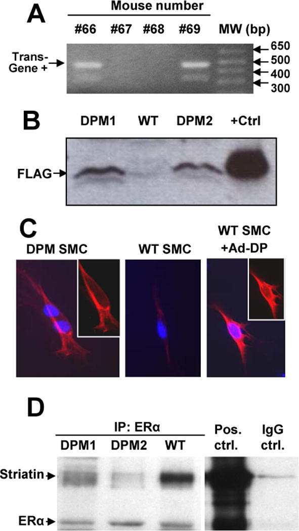
Interactions between ERα and striatin are blocked in a novel ERα176-253 Disrupting Peptide Mouse (DPM). (A) Tail DNA was amplified using primers flanking the ERα176-253-FLAG insert, yielding an ~480 bp transgene product – mice #66 and #69 were positive for the transgene. (B) The ERα176-253-FLAG peptide, as detected by anti-FLAG Western blot, is expressed in DPM but not WT liver cells. “+Ctrl”: Cos-1 cells transfected with an ERα176-253-FLAG expression plasmid. (C) Immunostaining for the disrupting peptide ERα176-253-FLAG (red) in mouse smooth muscle cells. Insets: same image without overlay of blue DAPI nuclear stain. (D) Lysates from liver samples of two different DPM mice, and one WT mouse were immunoprecipitated with anti-ERα antibody, before Western blotting with antibodies to Striatin or ERα. “Pos. ctrl”: positive control immunoprecipitation with both anti-ERα and anti-striatin antibodies from Cos-1 cells overexpressing ERα. “IgG ctrl.” Negative control immunoprecipitation with non-immune IgG antibody.