
Figure 6.
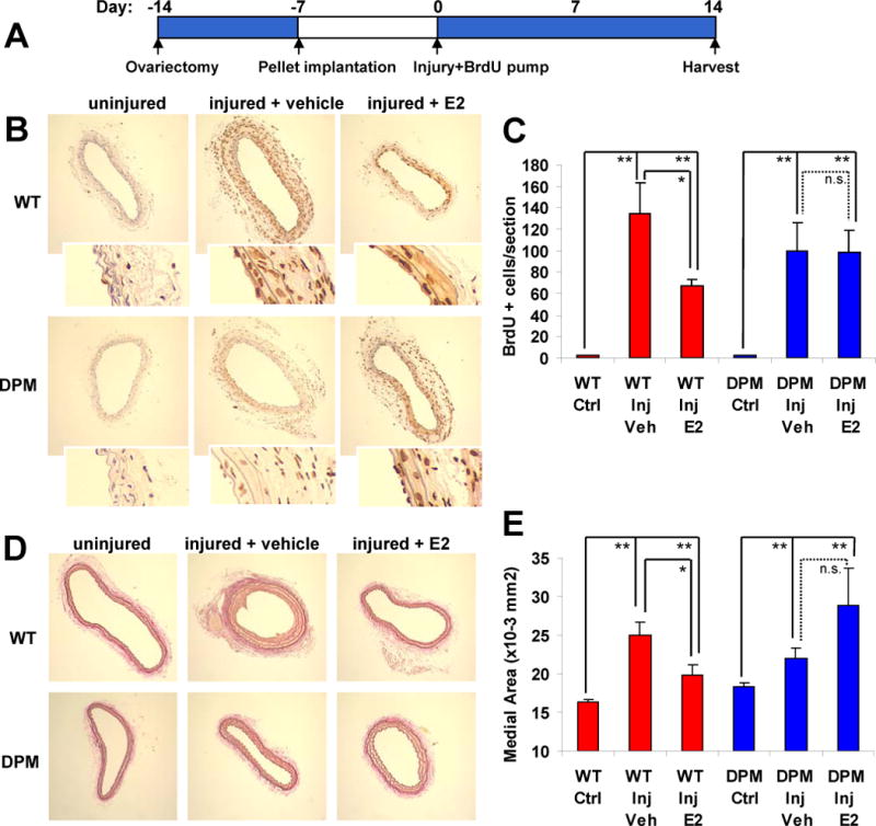
The ability of estrogen to protect against carotid arterial injury is lost in DPM mice. (A) Time course of the carotid artery wire injury model. 11 to 12 mice were used for each condition (see Supplemental Table 4 for weight measurements and other control parameters). (B) Representative anti-BrdU stained carotid artery sections, showing the effect of E2 on SMC proliferation in WT versus DPM mice. Insets: 4x higher magnification to show that BrdU stain is localized to cell nuclei. (C) Quantitation of BrdU positive cells per section. (D) Representative elastin stained sections showing the effect of estrogen on medial thickness in DPM versus WT mice. (E) Quantitation of medial thickness measurements. Bars: standard error of the mean. “*”: p<.05, “**”: p<.01.=, “n.s.”: no significant difference.