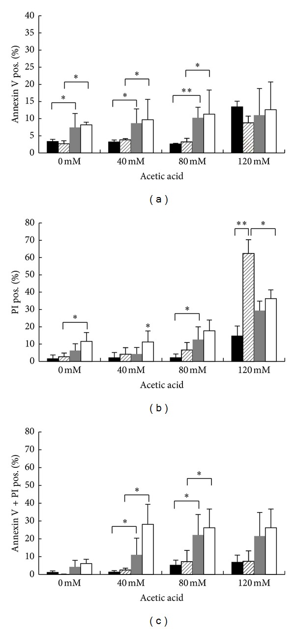
Figure 7.

Effect of YCA1 deletion on cell death after treatment with acetic acid. W303-1A (black bars), yca1Δ (black hatched bars), hxk2Δ (gray bars) and hxk2Δ-yca1Δ (white bars) exponentially growing cells were treated with different concentrations (40–80–120 mM) of acetic acid for 200 minutes at 30°C. Cell death was assessed by flow cytometry using FITC-coupled annexin V and PI co-staining to determinate the externalization of phosphatidylserine and the membrane integrity. The means of 3 independent experiments with standard deviations are reported. Student's t-test *P < 0.05 and **P < 0.01.
