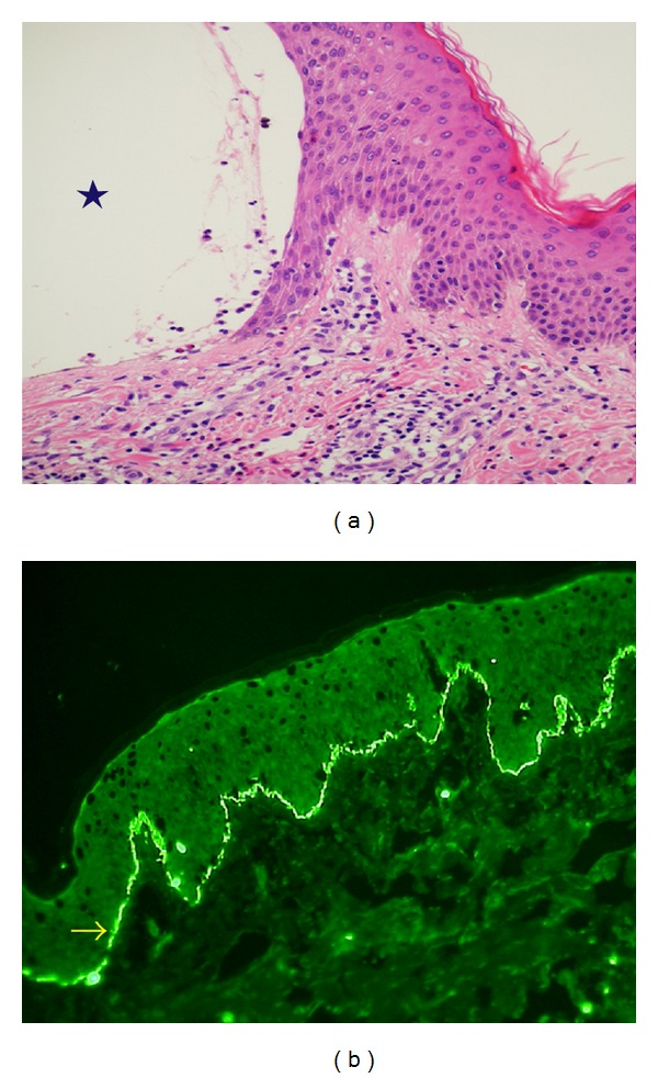
Figure 3.

Biopsy specimens of the skin lesions. (a) The hematoxylin and eosin staining shows subepidermal blister formation (as the star indicates) with cellular infiltration composed mainly of lymphocytes with numerous eosinophils. (b) Direct immunofluorescence shows linear complement C3 deposition (as the arrow indicates) along the basement membrane zone.
