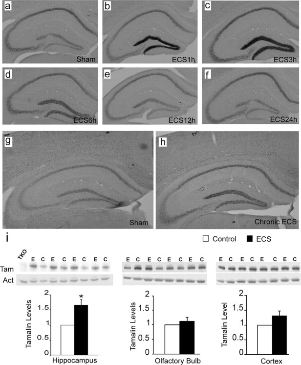
Figure 2.
Hippocampal tamalin mRNA and protein are upregulated in response to ECS. Representative tamalin in situ hybridization analysis of hippocampus from mice after single (a–f) or chronic (g, h) ECS. Sham-treated animals show the basal level of tamalin expression (a, g). Tamalin mRNA is upregulated at 1 h (b) and peaks at 3 h (c) from ECS. Beginning at 6 h (d), tamalin is downregulated and returns to the pre-ECS level by 12 (e) and 24 h (f). Note that tamalin mRNA expression remains upregulated at 24 h after chronic ECS (h) compared with sham-treated controls (g). n = 4 for each group. Expression of tamalin protein after chronic ECS (i). Animals received one ECS per day for 5 consecutive days and were killed 24 h after the last treatment. Western blot analysis of hippocampus (left), olfactory bulb (middle), and cortex (right) from ECS-treated (E) or sham-treated (C) WT animals using an anti-tamalin antibody (Tam); β-actin was used as loading control (Act), and a hippocampus lysate from a TKO mouse was used as negative control (TKO). The quantification of tamalin protein levels is shown at the bottom. Note the selective tamalin upregulation in the hippocampus. n = 4 animal. p < 0.05 by Student's t test.

