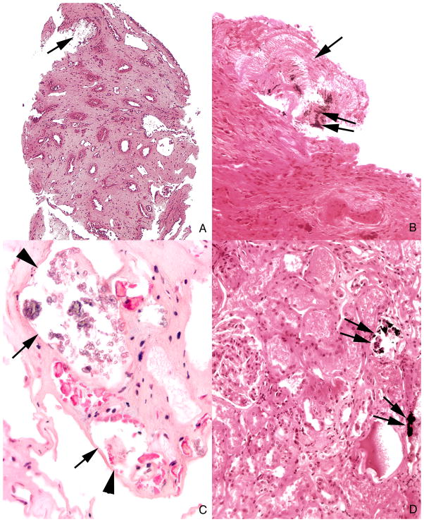
Figure 5. Histologic images of tubules with Yasue negative deposits in inner medulla and Yasue positive deposits in cortex.
Panels a and b each show a large deposit in a duct of Bellini (arrow) that is Yasue negative except for a very small region of Yasue positive staining (double arrow). These plugged ducts are generally very dilated, completely filled with mineral, surrounded by interstitial fibrosis and lack tubular lining cells. Several tubules filled with Yasue negative deposits (arrows) are seen at a higher magnification in panel c to show the loss of the tubular lining cells (arrowheads). Panel d shows two cortical collecting ducts stained with Yasue positive deposits (arrows). Magnification, x100 (a); x200 (b); x300 (c); x200 (d).

