Figure 4.
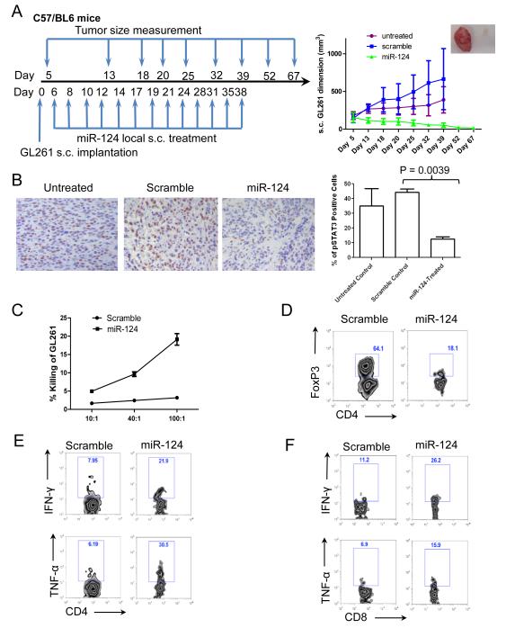
miR-124 exerts potent efficacy to suppress subcutaneous GL261 tumors in a syngeneic mouse model. (A) The treatment schema and the volumes of subcutaneous GL261 tumors in C57BL/6J mice treated intratumorally with either miR-124 or scramble control, or left untreated starting on day 6 (n=10/group/experiment). The figure is the result of a single experiment. In the miR-124 group, * denotes a P value of <0.01 compared with both the untreated and scramble control tumors. Standard deviations are shown. Arrows indicate days of treatment and tumor size measurements. The inset photo on the right is of a representative ex vivo GL261 tumor comparison between miR-124- and scramble control-treated tumor-bearing mice. (B) Ex vivo immunohistochemical analysis of gliomas, untreated (n=2), or treated with a scramble control (n=5), or miR-124 (n=3), which demonstrates a marked decrease in p-STAT3 expression in the local tumor microenvironment (P = 0.0039). (C) Splenocytes from miR-124 intratumorally treated GL261 mice (n=3) have markedly increased cytotoxicity at 48 hours after coculture with GL261 cells compared with that in splenocytes from scramble control oligonucleotide-treated tumor-bearing mice (P < 0.001). The ratios of splenocytes to GL261 cells are 10:1, 40:1, and 100:1. Error bars in the curve represent the standard deviation in the data from 3 mice. (D) Decreased tumor-infiltrating FoxP3+ Tregs in miR-124-treated GL261 mice. A single example is shown, but identical results were obtained in two other tumor bearing animals. Furthermore, miR-124 administration enhances IFN-γ and TNF-α production in CD4 T-cells (E) and CD8 T-cells (F) in the tumor local microenvironment. One set of representative FACS plots is shown. Similar results were obtained from another two tumor-bearing mice treated with miR-124 or scramble control, respectively.

