
Fig. 2.
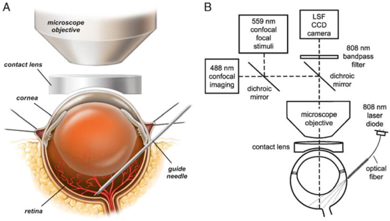
Drawings of the in vivo rat preparation. (a) The retina is imaged through an upright microscope and a contact lens placed over the cornea. A hypodermic needle is advanced through the sclera into the vitreous humor and serves as a guide needle through which a smaller needle is inserted to eject dyes onto the retinal surface. (b) The schematic shows the optical path for imaging the retina with confocal microscopy while simultaneously monitoring retinal blood flow with laser speckle flowmetry (LSF). The retina is illuminated for LSF by 808 nm light passing though an optical fiber that is pressed against the outer surface of the eye. A dichroic mirror directs the appropriate wavelengths of light from the retina to the LSF camera and the confocal microscope. An 808 nm bandpass filter prevents confocal excitation or emission light from entering the LSF camera. The confocal microscope has a primary scanner (488 nm) for imaging retinal vessels and a secondary scanner for generating 559 nm stimulating light flashes. The secondary scanner also generates 405 nm light pulses for photolysis of caged compounds. See ref. (12) for a further discussion of LSF. Modified from ref. (12).