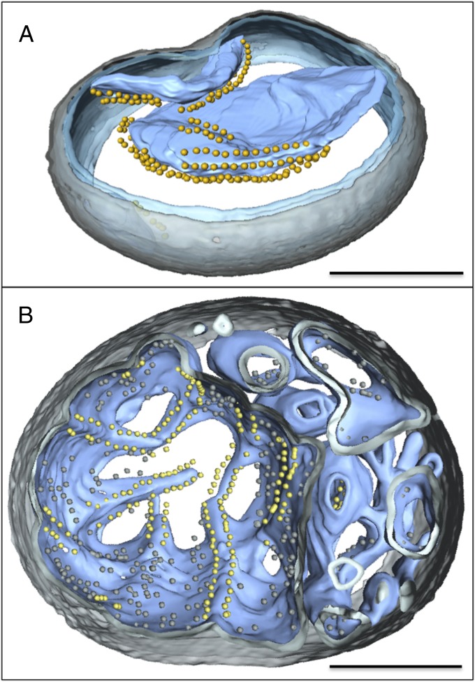
Fig. 2.
Three-dimensional tomographic volumes of mitochondria with standard or early-vesicular morphology. Segmented tomographic volume of mitochondria with standard (A) and early-vesicular morphology (B). In A, the ATP synthase dimers are arranged in rows along highly curved apices of inner-membrane cristae. In B, ATP synthases dimer rows are arranged along shallow inner-membrane ridges in matrix compartments with diameters >350 nm. Smaller inner-membrane vesicles show no evidence of ATP synthase dimers or dimer rows. Outer membrane, transparent gray; inner membrane, transparent blue; cristae, light blue. ATP synthase F1 heads are shown as yellow spheres. (Scale bars, 200 nm.)

