
Fig. 2.
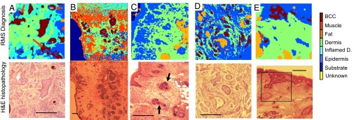
Diagnosis of BCC by Raman microspectroscopy using raster scanning. Tissue sections: (A and B) nodular BCC; (C) BCC-negative skin sample containing hair follicles with morphology similar to that of BCC (black arrows). Unsectioned tissue layers: (D) infiltrative BCC; (E) superficial BCC. The histopathology images for adjacent sections are included for comparison. (Scale bars: 400 μm.) False positives are indicated by red arrows.