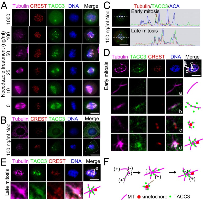
Fig. 4.
Targeting of preassembled acentrosomal TACC3–microtubule seeds/asters to kinetochore facilitates kinetochore capture by centrosomal microtubules. (A) TACC3–microtubule complex dynamically associated with kinetochores upon nocodazole treatment. Staining of mitotic HeLa cells arrested by different concentrations (1 μg/mL, 100 ng/mL, 50 ng/mL, 25 ng/mL, 10 ng/mL, and 0 ng/mL) of nocodazole. Tubulin is in magenta, CREST in red, TACC3 in green, and DNA in blue. (B) The total of 100 ng/mL nocodazole-arrested HeLa cells in early mitosis were stained with tubulin (magenta), CREST (red), TACC3 (green), and DNA (blue). The white dashed lines indicate the cell boundary. (C) Illustration of acentrosomal TACC3–microtubule (MT) seeds/asters in 100 ng/mL nocodazole-arrested early and late mitotic HeLa cells. (D and E) Different types of acentrosomal microtubules assemble the mitotic spindle. HeLa cells were treated with 25 ng/mL nocodazole for 5 h. The representative early (D) and late (E) mitotic cells are shown. The maximum intensity projections of 3D images are shown. In early mitosis (D), four different types of acentrosomal microtubule structures (a–d) are indicated by projection of selected z stacks followed by magnification. In contrast, only one type of acentrosomal microtubules (e in E) was in late mitosis. The square e in E is magnified and illustrated (Below). (F) Illustration of the acentrosomal microtubule nucleation, TACC3–microtubule seeds/asters assembly and the end-on capture of the kinetochores by the small aster tubules during transition from early mitosis to late mitosis. (Scale bar, 10 μm.)

