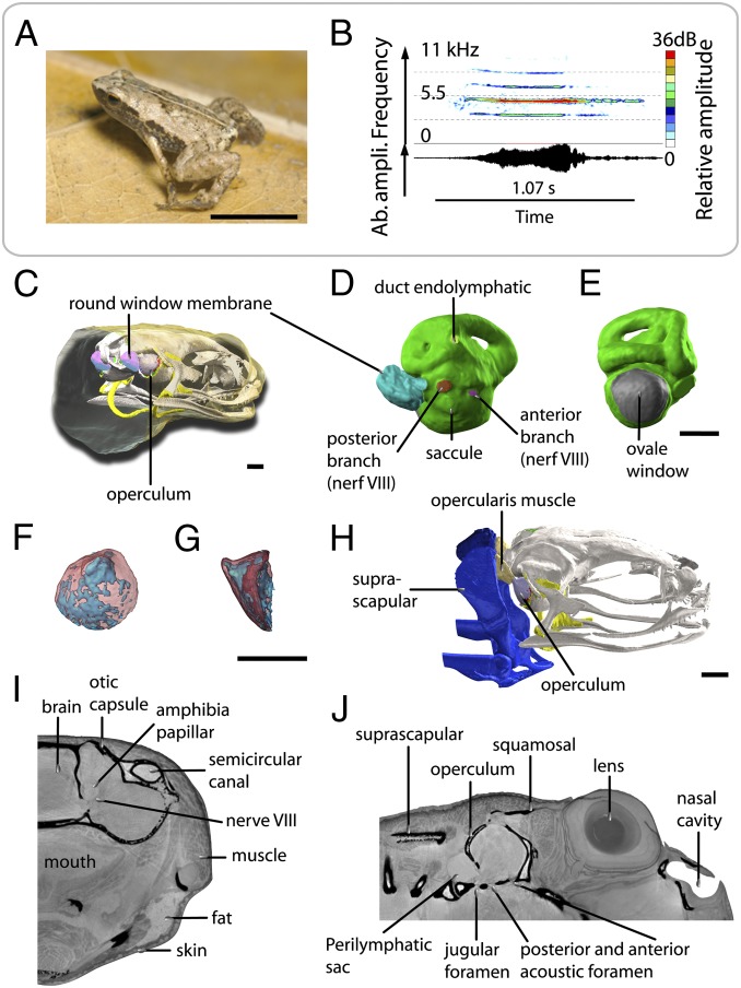
Fig. 1.
Acoustic behavior and volume rendering of ear region of S. gardineri. (A) Picture of one adult male of S. gardineri. (B) Oscillogram (Lower) and sonogram (Upper) of the advertisement call. (C) 3D visualization of the operculum and endolymphatic sac of the oval window in the head, in 3/4 posterior view with the skin made transparent. (D) Median view of the inner ear (in green) with the endolymphatic sac connected to the left round window, showing the location of the posterior (red) and anterior (violet) branches of nerve VIII, the endolymphatic duct (pale green), and extracranial lymphatic sac (blue-green). (E) Posterior view of the left inner ear, showing the surface occupied by the oval window (in gray). (F and G) 3D visualization of the operculum in transparency (bone in red, cartilage in blue): posterior (F) and lateral (G) views. (H) 3D 3/4 lateral view of the opercular system with the opercular muscle shown in pink. (I and J) Sagittal and frontal sections of holotomographic images showing the foramen of the otic capsule, the innervation of the inner ear, and the organization of the tissues surrounding the ear. (Scale bar, 0.5 mm.)

