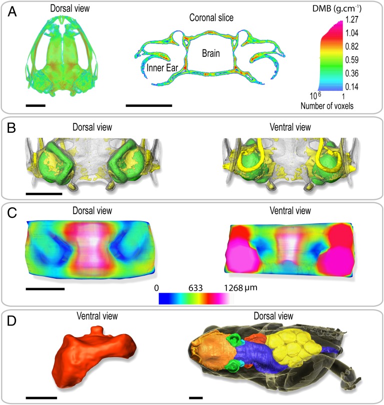
Fig. 2.
Volume rendering of holotomography of the body and the tissues surrounding the ear of S. gardineri. (A) 3D map (Left) and virtual sagittal section (Right) illustrating the degree of mineralization of the skull as determined by absorption tomography. (B) Visualization of the degree of ossification of the otic region of the skull in dorsal (Left) and ventral (Right) views. The skull is transparent, cartilage is shown in yellow, and the inner ears in green. (C) 3D map of the thickness of the tissues separating the inner ears and the gaseous medium. (Left) Dorsal view and (Right) ventral view of the oral region. The thinnest parts are shown in dark blue. (D) 3D visualization of the pulmonary system of a gravid female (SVL = 11.65 mm). Red, pulmonary and laryngeal cavity in ventral view. To right, dorsal view of female in transparency visualizing the anatomy and the volume occupied by the nasal and oral cavities (orange), the inner ears (green), the digestive system (blue), the ovarian masses (yellow), and the pulmonary system. Note the left forelimb was removed for molecular analysis. (Scale bar, 1 mm.)

