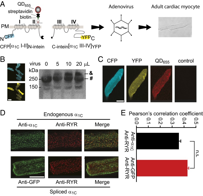
Fig. 4.
Expression and subcellular targeting of intein-spliced α1C in cardiac myocytes. (A) Schematic showing incorporation of split-intein–tagged α1C moieties into adenovirus and infection of cardiomyocytes. (B, Left) Confocal images showing high efficiency expression of CFP- and YFP-tagged split-intein α1C moieties in adult cardiomyocytes. (Scale bar, 50 μm.) (B, Right) Western blot (anti-α1C antibody) detection of endogenous (#) and intein-spliced (&) α1C subunits. (C) Confocal images of CFP, YFP, and QD655 fluorescence in a cardiomyocyte expressing intein-spliced α1C. Control shows absence of QD655 staining in an uninfected cell. (Scale bar, 20 μm.) (D, Upper) Immunostaining of endogenous α1C (anti-α1C, green) and RyR (anti-RyR, red) in an uninfected cardiomyocyte. (D, Lower) Immunostaining of intein-spliced α1C (anti-GFP, green) and endogenous RYR (anti-RyR, red). (Scale bar, 20 μm.) (E) Colocalization analyses.

