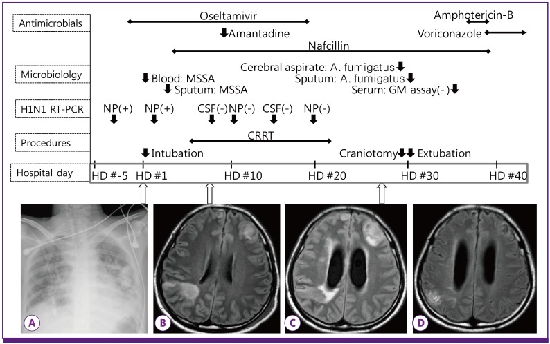
Figure 1.
Sequence of clinical and laboratory events. Reverse transcription-polymerase chain reaction for H1N1 virus was performed serially by nasopharyngeal swab (HD #-3, 2, 10, and 19) and CSF (HD #8 and 14). Initial chest radiography (A) showed multiple patchy consolidations. MRI showed a vague rim-enhancing mass lesion on HD #8 (B), and the abscess became evident on HD #27 (C). The lesion disappeared after surgery and 18 weeks of anti-fungal therapy (D).
MSSA, methicillin-sensitive Staphylococcus aureus ; GM, galactomannan; NP, nasopharynx; CSF, cerebrospinal fluid; CRRT, continuous renal replacement therapy; HD, hospital day. HD #1 indicates the day of admission.

