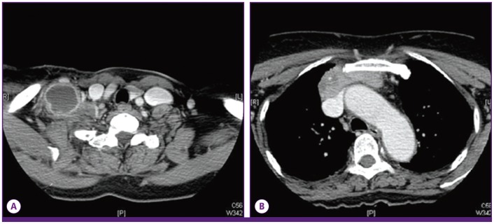
Figure 1.
(A) The chest computed tomography scan shows an enlarged lymph node with necrosis in the right supraclavicular area and enhancement of the surrounding area. (B) The chest computed tomography scan shows a mass in the anterior mediastinum with necrosis that runs continuously from the supraclavicular area to the anterior mediastinum.

