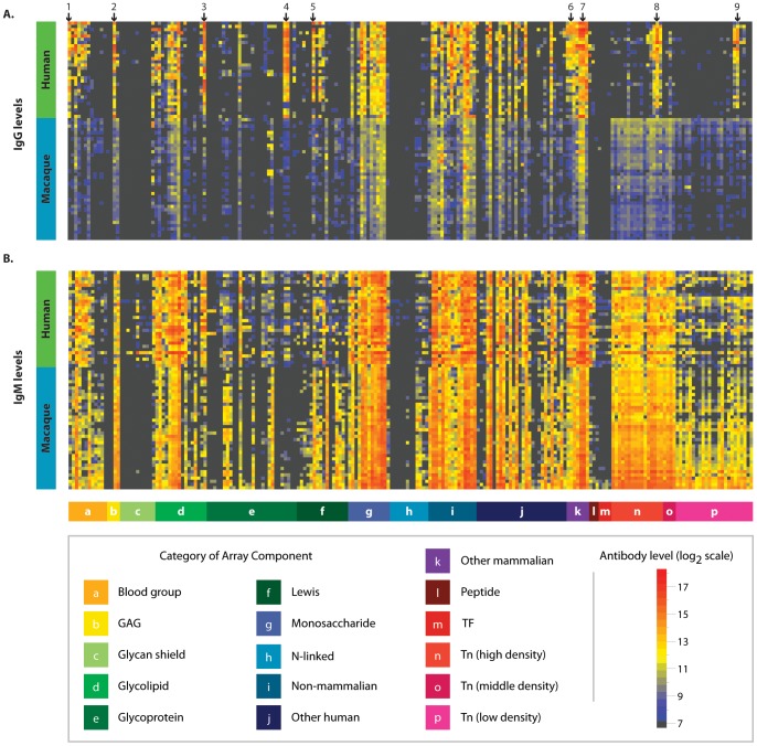
Figure 2. Comparison of anti-glycan antibody profiles in macaques and humans.
Heat map showing glycan binding of circulating IgG (A) and IgM (B) in pre-vaccinated macaques (n = 38) and healthy humans (n = 30). Columns correspond to individual glycans organized into glycan families (see legend). Rows represent individual humans and macaques, which have been sorted by hierarchical clustering. Macaques and humans have highly similar repertoires of anti-glycan antibodies. Highlighted glycans are (1) 2′FucLac, (2) Hya8, (3) P1, (4) Ovalbumin, (5) LeA, (6) alpha-gal, (7) Forssman di, (8) Ac-Tn(Thr)-G-21, and (9) Ac-Tn(Thr)-Tn(Thr)-Tn(Thr)-G. Normalized data are plotted on a log 2 scale with a floor value of 7.2 (colored black).

