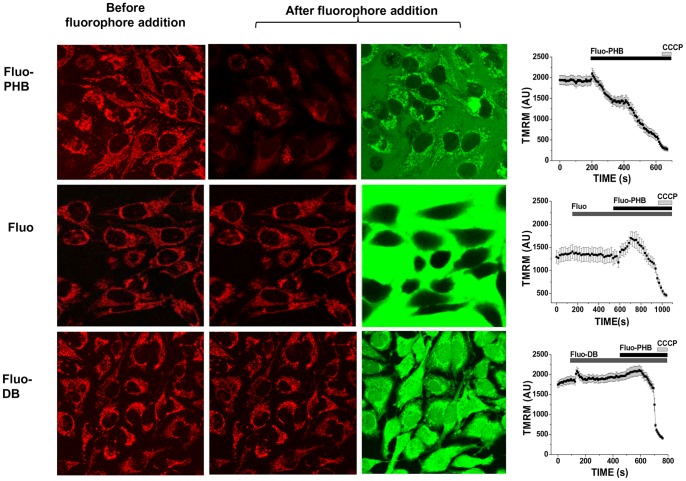
Figure 3. Fluo-PHB but not fluorescein or fluo-DB induces mitochondrial membrane depolarization.
HeLa cell loaded with 25/ml of fluorescent probes and imaged with a confocal microscope over time. Left column shows TMRM fluorescence in mitochondria before treatment. Second and third columns show TMRM and fluorescein after addition of the probes. Fluorescein did not distribute inside the cells, while fluo-DB did not show preferential mitochondrial localization. Note that neither fluorescein nor fluo-DB affected mitochondrial membrane potential, which was decreased only in the presence of fluo-PHB. This is represented in the graphs that are in the left column that show TMRM fluorescence in arbitrary units (AU) collected from the mitochondrial regions of the intact cells as a function of time. Scale bar 20 µm.

