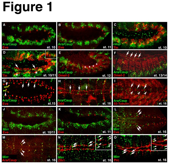
Figure 1. Expression pattern of Ara/Caup and Mirr during embryogenesis in wild-type embryos.
(A) Double immunostaining for Ara/Caup and Eve shows the mesodermal expression of Ara/Caup as a continuous band along the dorsal side of the embryo. (B) During stage 11 Ara/Caup proteins are present in segmental clusters abutting the Eve cell clusters. (C) In situ hybridization for caup transcripts and immunostaining for Tin further demonstrates the presence of caup in the dorsal mesoderm. (D) Starting at stage 11 Tin-positive cells segregate into cardiac cells (arrows) and cells of the visceral mesoderm (asterisks). There is still a considerable overlap of Tin and caup mRNA expression. The inset in (D) show a higher magnification to better visualize caup mRNA transcripts around Tin protein that is located in the nucleus. (E) By stage 12 Ara/Caup expression has vanished from the dorsal mesoderm (asterisks) where heart cells begin to differentiate. (F) Starting at mid-embryogenesis, single Ara/Caup-positive cells arise along the Dmef2 expressing myocardial cell row (arrows). (G) Double labeling for Ara/Caup and Eve at stage 15 shows their co-expression in the most anterior Eve-positive pericardial cells (arrowheads), as well as Ara/Caup-only expressing cells located between the Eve pericardial cells (arrows). (H) The Ara/Caup-expressing cells (arrows) are not positive for the pericardial cell marker Odd. (I) β3-Tubulin that labels the four Tin-positive myocardial cells in each hemisegment by stage16 and allows a more accurate localization of the Ara/Caup expressing cells (arrows) along the heart tube. The arrowheads point to the β3-Tubulin-negative myocardial cells in each segment. These cells express Seven-up and Doc. (J) Mirr expression in the dorsal mesoderm is similar to the expression of Ara/Caup at early embryonic stages. (K) During stage 11, Mirr protein accumulates around the Eve-positive cell clusters. (L) At stage 16, pairs of Mirr-only expressing cells are detected between the Eve pericardial cells (arrows). (M) Double labeling for Ara/Caup and Mirr reveals the co-expression of all three factors in one cell (arrowheads) of the segmentally arranged Mirr expressing (arrows) cell pairs. (N) None of the Zfh-1-expressing pericardial cells co-expresses Mirr. (O) Double immunostaining for Mirr (arrows) and the extracellular matrix molecule Pericardin (Prc) that labels pericardial cells and is expressed along the extensions of the alary muscles, shows that the Mirr-positive cells are located between these extensions. Additionally, one of the two Mirr-positive cells lies adjacent to the Prc-expressing basal membrane of the myocardial cells. All embryos are oriented with the anterior to the left. Embryos in A-F, J, K are shown from the lateral side. A dorsal view of the embryos in G, H, I, L-O is shown.

