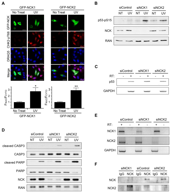
Figure 5. Specific contributions of the NCK isoforms.
(A) HeLa cells transfected with GFP-NCK1 or GFP-NCK2 were treated with 50 J/m2 UV and allowed to recover for 2 hr before being fixed and stained with indicated antibodies and DRAQ5. Scale bars are 20 µm. All images are confocal sections. Bar graphs below each panel are ratio of nuclear to cytoplasmic fluorescence of GFP-NCK with no treat defined as 1. n = 14-28; error bars represent SE; (*) P < 0.001, (**) P < 0.0001. (B) HeLa cells transfected with control, NCK1, or NCK2 siRNA were treated with 50 J/m2 UV and allowed to recover for 2 hr before lysates were prepared. Equal amounts of lysates were immunoblotted for p53-pS15 (phospho-specific) and NCK. RAN was used as a loading control. n = 3. (C) RT-PCR of p53 mRNA from HeLa cells transfected with control, NCK1, or NCK2 siRNA. GAPDH was used as an internal control. RT = reverse transcriptase. n = 4. (D) HeLa cells transfected with control, NCK1, or NCK2 siRNA were treated with 50 J/m2 UV and allowed to recover for 2 hr before lysates were prepared. Equal amounts of lysates were immunoblotted for cleaved CASP3, total CASP3, cleaved PARP, total PARP, and NCK. RAN was used as a loading control. n = 3. (E) RT-PCR of NCK1 and NCK2 mRNA from HeLa cells transfected with control, NCK1, or NCK2 siRNA. GAPDH was used as an internal control. n = 3. (F) Cell lysates from HeLa cells transfected with control, NCK1, or NCK2 siRNA were immunoprecipitated with a NCK antibody, which detects NCK 1 and NCK2, or IgG control, and immunoblotted for NCK or NCK2. n = 4.

