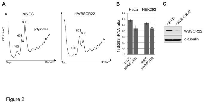
Figure 2. Polysome analysis of HeLa cells after depletion of WBSCR22.
(A) HeLa cells were transfected with siWBSCR22 and siNeg, the cytoplasmic cell extracts were prepared 72 h after transfection and centrifuged on 10-50% sucrose gradient. Absorbance at 254 nm was measured across the gradient, and the positions corresponding to the 40S, 60S and 80S ribosomal particles are indicated. (B) The ratio of 18S/28S rRNA from HeLa and HEK293 cells transfected with siRNAs. RNA was purified from cytoplasmic extracts of siWBSCR22 and siNeg cells, separated by electrophoresis and intensities of 18S and 28S rRNA were quantified. The P value is 0.04 using Student’s t-test. (C) Protein expression of siWBSCR22 and siNeg. transfected cells was determined by western blot analysis using anti-WBSCR22 and anti-tubulin antibodies.

