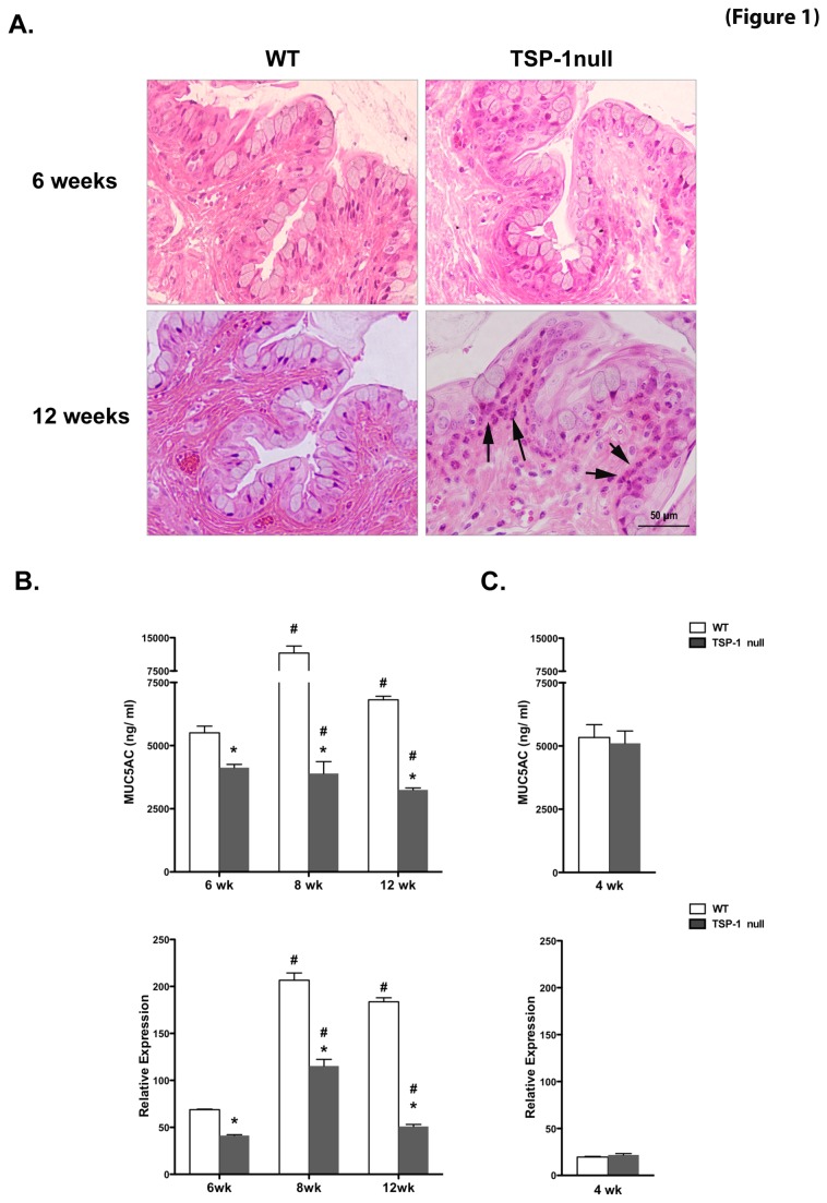
Figure 1. Inflammatory damage and diminished mucin levels are detectable in TSP-1 null conjunctiva.
(A) Histology of conjunctiva from WT and TSP-1 null mice at 6 and 12 weeks. Representative hematoxylin-eosin stained sections at 6 weeks show normal epithelial morphology and a lack of abnormal inflammatory cells. At 12 weeks, TSP-1 null conjunctiva displayed prominent inflammatory infiltrates (arrows) in the epithelial layer adjacent to goblet cells. (B-C) Pilocarpine-induced tears and conjunctival tissues collected from WT or TSP-1 null mice (n=5 each) were analyzed for levels of MUC5AC using ELISA and real time PCR, respectively. At 6, 8, and 12 weeks of age, goblet cell-derived tear mucin content and conjunctival message were significantly diminished in TSP-1 null mice compared to age-matched controls, while no significant differences were detected at 4 weeks of age. Results are presented as ng/ml for ELISA assay and as relative expression to that of housekeeping gene GAPDH for real time PCR (*p < 0.05 compared to WT control; #p < 0.05 compared to 4 weeks).

