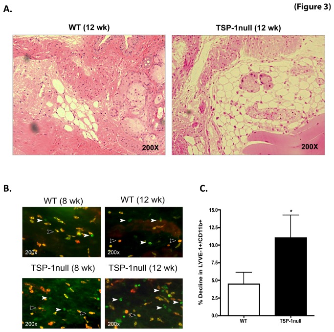
Figure 3. Increased lymphatic vessels in TSP-1 null conjunctiva are accompanied by a decline in LYVE-1+ macrophages.
(A) Hematoxylin and eosin stained sections of the conjunctiva harvested from 12 week old WT or TSP-1 null mice show relatively increased lymphatic vessels in TSP-1 null conjunctiva. (B) Whole mounts of conjunctiva from 8 and 12 week old WT and TSP-1 null mice were immunostained with fluorochrome conjugated anti-LYVE-1(red) and anti-CD11b (green) antibodies. (C) Quantitative analysis of positively stained cells showed significantly increased decline in LYVE-1 expressing CD11b+ macrophages at 12 week (from 8 week) of age in TSP-1 null mice as compared to WT mice (* p < 0.05, compared to WT)..

