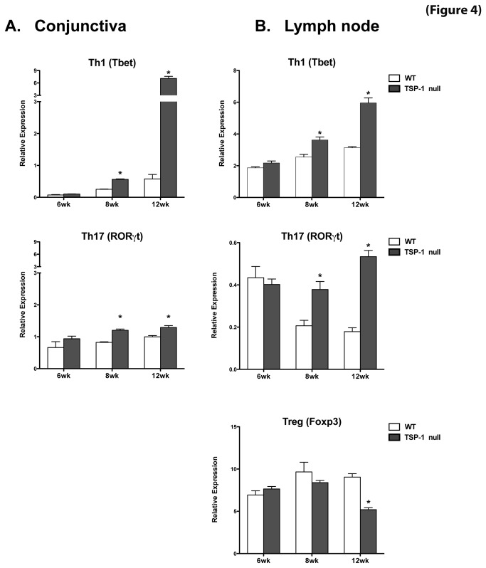
Figure 4. Inflammatory effectors are detected in the conjunctiva and the draining lymph nodes of TSP-1 null mice.
Conjunctiva tissue and cervical lymph node cells were collected from WT and TSP-1 null mice at 6, 8, and 12 weeks (n=3 each). Extracted RNA was analyzed in a real-time PCR assay to determine the levels of message for the transcription factors Tbet, RORγt and Foxp3. No significant differences were detected in Tbet or RORγt expression in conjunctiva (A) and lymph nodes (B) at 6 weeks of age, while at 8 and 12 weeks the expression of both transcription factors was significantly increased in TSP-1 null mice compared with the WT control tissues. Expression of Foxp3 was significantly decreased in TSP-1 null lymph nodes at 12 weeks of age. Results are presented as relative expression of a transcription factor gene to that of the housekeeping gene GAPDH (* p <0.05 as compared to WT controls)..

