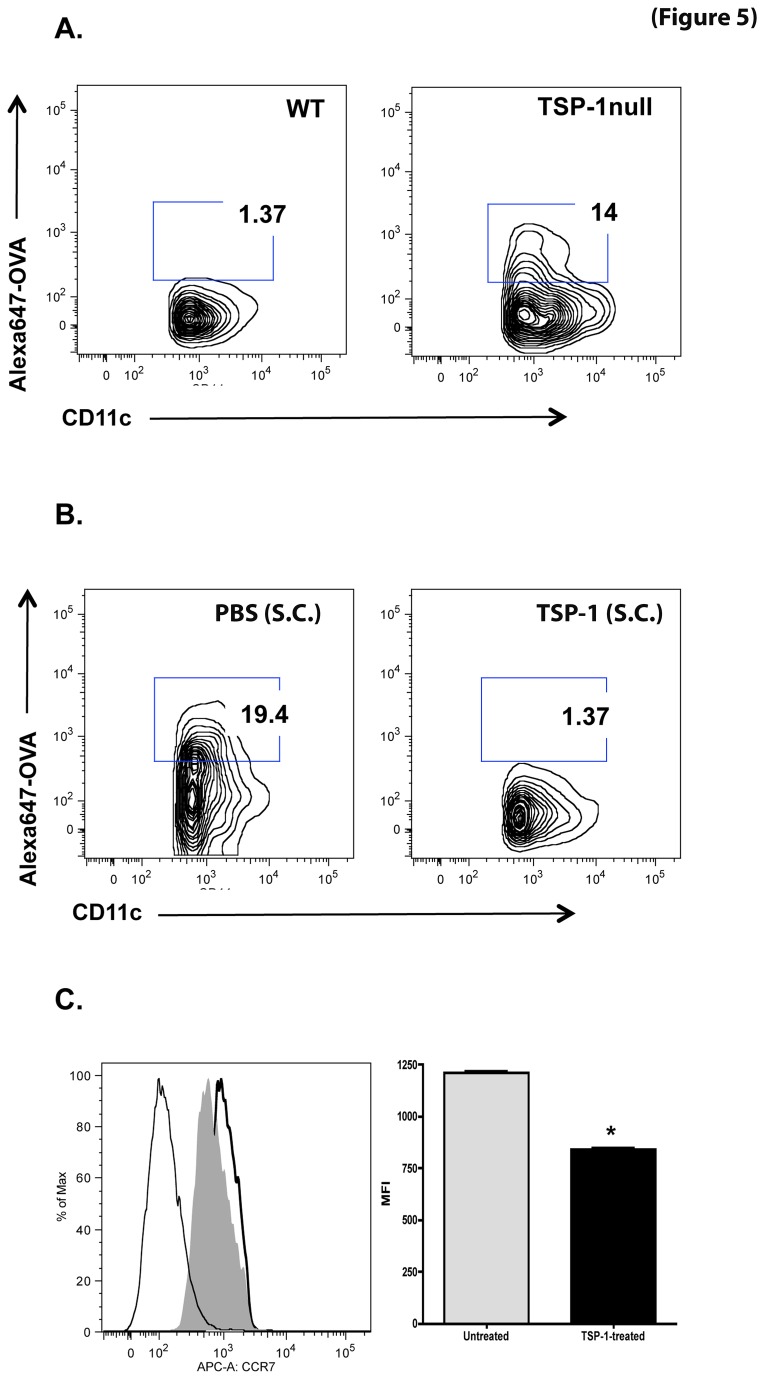
Figure 5. Migration of DCs carrying ocular surface antigens to draining LNs is enhanced in TSP-1 null mice.
Cervical lymph nodes from 10-week old WT and TSP-1 null mice (n=3 each) were harvested 3 hr after topical application of Alexa647-conjugated OVA (125 µg/2.5 µl per eye). Lymph node cells were stained with fluorochrome-conjugated CD11c prior to analysis by flow cytometry and CD11c/Alexa647+ cells were identified. (A) A marked increase in CD11c/Alexa647+ cells was detectable in TSP-1 null lymph node cells compared to WT controls. (B) Blockade of CD11c/Alexa647+ dendritic cell migration to draining lymph nodes was observed in TSP-1 null mice following subconjunctival (S.C) injection of TSP-1 as compared to PBS-injected control group. (C) BMDC were treated with TSP-1 for 24 hr, stained with fluorochrome-conjugated CCR7, and analyzed by flow cytometry. The histogram shows fluorescence detected in unstained (thin line), untreated (thick line) and TSP-treated (filled grey) cells. The bar graph shows mean fluorescence intensity (MFI) of CCR7 staining. TSP-1 treatment significantly decreased the expression of CCR7 (* p<0.05).

