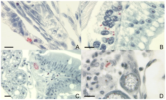
Figure 1. Spores of Pseudoloma neurophilia in Luna-stained histological sections of progeny of infected zebrafish, Danio rerio.
A.Spores (red) in the epidermis of a 7 d post-fertilization (pf) larval zebrafish. B. Spores in the resorbing yolk-sac of the same 7 dpf larval zebrafish. C. Spore aggregate beneath the intestinal epithelium of an 8 wk pf juvenile fish. D. Spores in the ovigerous stroma adjacent to developing follicles in an 8 wk pf fish. Bar = 10 µm.

