Figure 15.

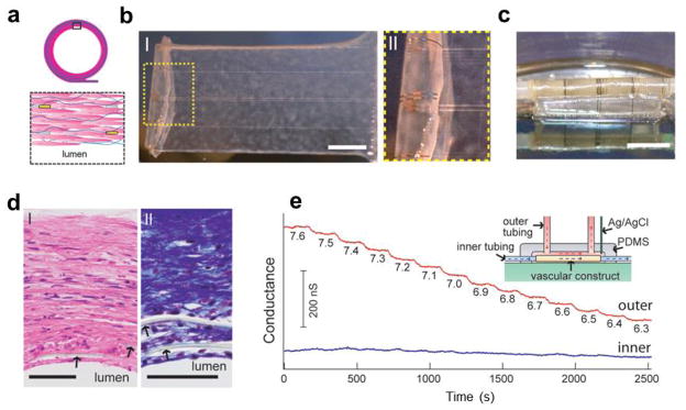
Synthetic vascular construct enabled 3D sensing. (a) Schematic of the smooth muscle nanoES. The upper panels are the side view, and the lower one is a zoom-in view. Grey: mesh nanoES; blue fibers: collagenous matrix secreted by HASMCs; yellow dots: nanowire FETs; pink: HASMCs; (b) (I) Photograph of a single HASMC sheet cultured with sodium L-ascorbate on a nanoES. (II) Zoomed-in view of the dashed area in I. Scale bar, 5 mm; (c) Photograph of the vascular construct after rolling into a tube and maturation in a culture chamber for three weeks. Scale bar, 5 mm; (d) Haematoxylin-Eosin- (I) and Masson-Trichrome- (II; collagen is blue) stained sections (~6 μm thick) cut perpendicular to the tube axis; lumen regions are labelled. The arrows mark the positions of SU-8 ribbons of the nanoES. Scale bars, 50 μm; (e) Changes in conductance over time for two nanowire FET devices located in the outermost (red) and innermost (blue) layers. The inset shows a schematic of the experimental set-up. Outer tubing delivered bathing solutions with varying pH (red dashed lines and arrows); inner tubing delivered solutions with fixed pH (blue dashed lines and arrows). Reprinted with permission from Ref. [39]. Copyright 2012 Nature Publishing Group.
