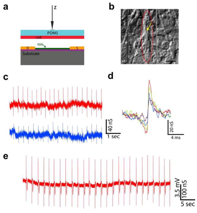
Figure 4.
Building interface between SiNW FETs and spontaneously firing cardiomyocytes. (a) Schematic illustration of manipulating the PDMS/cell substrate to make the interface with an NWFET device. (b) Distinct patch of beating cells (red dashed oval) over a SiNW device (yellow arrow). scale bar is 20 μm; (c) Two representative traces recorded with same SiNW FET from a spontaneously firing cardiomyocyte with different PDMS/cell displacement value; (d) High resolution comparison of single peaks recorded with increasing displacement values (from purple to red); (e) Data recorded in distinct experiment at displacement value close to cell failure. Reprinted with permission from Ref. [51]. Copyright 2009 National Academy of Sciences.

