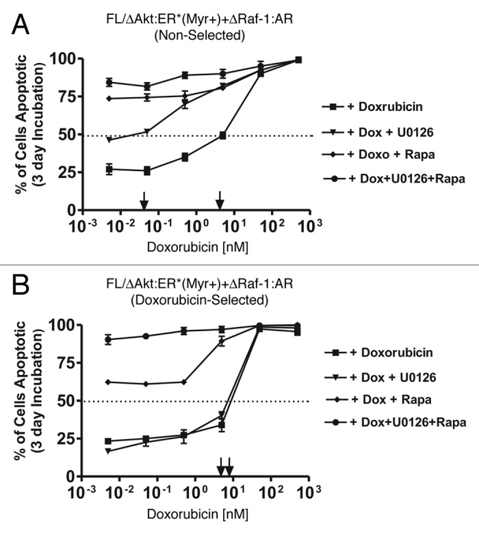
Figure 9. Effects of MEK and mTOR inhibitors on the induction of apoptosis induced by doxorubicin. The effects of MEK1 and mTOR inhibitors on doxorubicin IC50 was examined by culturing the indicated cells in the presence of the different concentrations of doxorubicin for three days and then determining the extent of apoptosis induction by annexin V/PI staining. Cells were cultured in medium containing 500 nM 4HT + 100 nM Test and the indicated concentrations of the inhibitors. (A) non-selected FL/ΔAkt-1:ER*(Myr+) + ΔRaf-1:AR cells, and (B) doxorubicin-selected FL/ΔAkt-1:ER*(Myr+) + ΔRaf-1:AR cells. Symbols: solid squares (■) = doxorubicin, solid downward triangles (▼) = indicated concentrations of doxorubicin and 5,000, 500, 50, 5, 0.5 and 0.05 nM MEK inhibitor, solid diamonds (◆) = indicated concentrations of doxorubicin and 500, 50, 5, 0.5, 0.05 and 0.005 nM mTOR inhibitors, and solid circles (●) = indicated concentrations of doxorubicin and 5,000, 500, 50, 5, 0.5 and 0.05 nM MEK and mTOR inhibitors.
