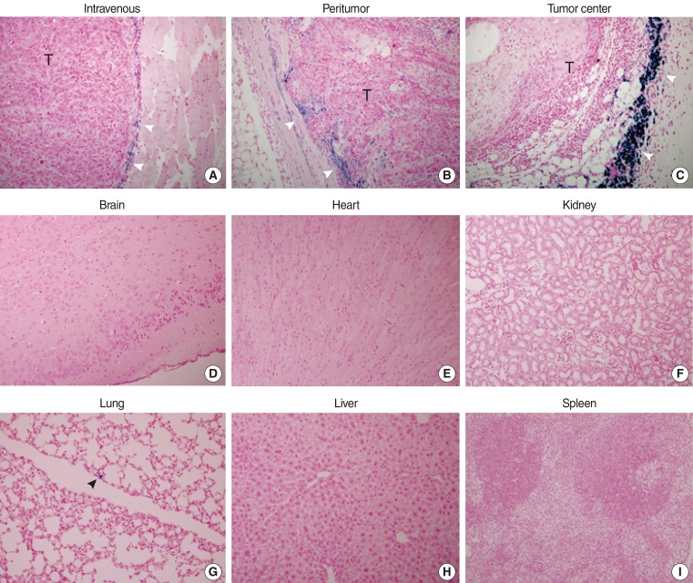
Fig. 3.
Tumor homing capabilities of HB1.F3-CD (F3-CD) cells (Prussian blue staining, ×40). F3-CDs were labeled with Feridex and injected into the tail vein (A), subcutaneously 1.5 cm from the tumor injection site (B), or at the tumor center (C) on 7 day after tumor cell inoculation. Mice were sacrificed on day 14, and Prussian blue staining was performed. F3-CDs were found primarily at the tumor border (white arrowheads). F3-CDs were not observed in tissues from brain (D), heart (E), kidney (F), liver (H), or spleen (I). F3-CDs were observed in lung tissue (G, black arrowhead) at a very low frequency.

