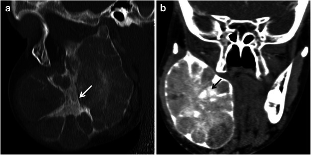
Fig. 10.

A 15-year-old boy with cemento-ossifying fibroma of the mandibular ramus. a Sagittal and (b) coronal reformatted images demonstrating a well-defined expansile lesion in the mandibular ramus with internal sclerotic bands (arrow) and some ground-glass matrix
