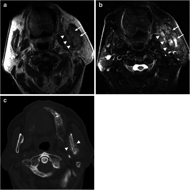
Fig. 12.

Lung cancer metastases in the masticator space. Axial (a) T1-weighted and (b) T2-weighted MR images show a heterogeneous mass centred in the left ramus of the mandible (arrowheads) with a soft tissue component (arrows). c Axial bone window CT scan at the same level shows sclerosis of the mandibular ramus and periosteal reaction in both cortices (arrowheads), suggestive of a malignancy
