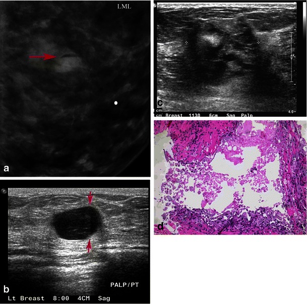
Fig. 10.

Galactocele: a 35-year-old lactating woman, presenting with a palpable lump. a Photographic magnification of left lateral mammogram shows a well-circumscribed lesion with a fat fluid level on the lateral projection. This is the classic appearance of galactocele. b Grey-scale ultrasound shows a cystic lesion which, on adjusting the gain, shows a fat/fluid level diagnostic of galactocele. c Another patient, a 38-year-old lactating woman, presenting with breast pain and palpable mass. Mammogram showed dense breast tissue with a partially obscured mass and skin thickening (not shown here). Grey-scale ultrasound image shows heterogeneous ill-defined mass, which was vascular on power Doppler. Biopsy was performed. d Microscopic high-power image (40×), revealing a ruptured galactocele with cyst contents leaking into the surrounding tissue, causing a lipogranulomatous inflammation and foamy histiocyte aggregation
