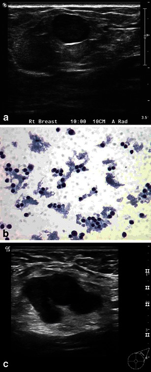
Fig. 12.

a Benign reactive lymph node: a 30-year-old pregnant woman, presenting with a palpable lump. Grey-scale ultrasound image shows well circumscribed hypoechoic nodule, with significant vascularity on power Doppler. b Fine-needle aspiration cytology reveals polymorphous population of lymphocytes and macrophages, all features of benign reactive lymph node. c Metastatic lymph node: a 39-year-old woman, 12 weeks pregnant, presenting with palpable axillary mass. The patient had a remote history of breast cancer treated with mastectomy. Gray-scale ultrasound showed an enlarged axillary node with abnormally thickened cortex. Core biopsy revealed metastatic breast cancer
