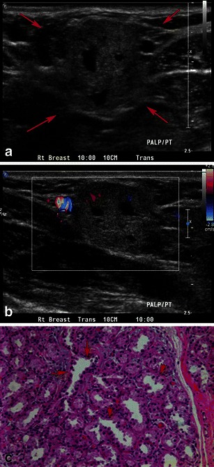
Fig. 5.

Lactational change: a 31-year-old lactating woman, presenting with lump in the right breast. a Grey-scale ultrasound shows a partially circumscribed hypoechoic nodule with cystic areas, (b) with internal vascularity on power Doppler. c Pathology slide at high power (20×) shows lobular expansion containing increased numbers of acini, many of which are enlarged and dilated, consistent with lactational change
