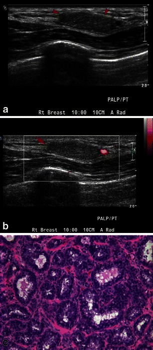
Fig. 6.

Lactating adenoma: a 38-year-old pregnant woman, presenting with a breast lump. a Grey-scale ultrasound image shows an isoechoic, circumscribed nodule. b Power Doppler image shows minimal peripheral vascularity. Biopsy was performed revealing lactating adenoma. c Higher-power image (20×) shows epithelial cell enlargement, cytoplasmic vacuolisation and a hobnail appearance with protrusion of cells into the acinar lumen
