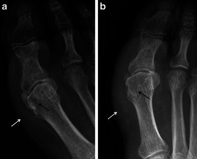
Fig. 12.

Osteomyelitis. a AP radiograph in a patient with a plantar ulcer showing cortical dehiscence and destructive change at the medial sesamoid (black arrow) suggestive of osteomyelitis. Note severe adjacent soft tissue swelling (white arrow). b AP radiograph in the same patient approximately 5 weeks later with near complete destruction of the medial hallucal sesamoid (black arrow) and persistent soft tissue swelling (white arrow). The great toe was amputated
