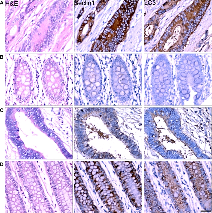
Fig. 1.
Expression of Beclin 1 and LC3 in colorectal cancer tissues. Immunohistochemical staining for Beclin 1 and LC3 were performed in paraffin-embedded sections of colorectal cancer and normal adjacent tissues. Most tumors showed Beclin 1 and LC3 with strong staining (a) in comparison to paired normal adjacent tissues (b). But some colorectal cancer tissues showed weak staining (c) in comparison to paired normal adjacent tissues (d). Representative pictures were presented. Scale bars, 50 μm

