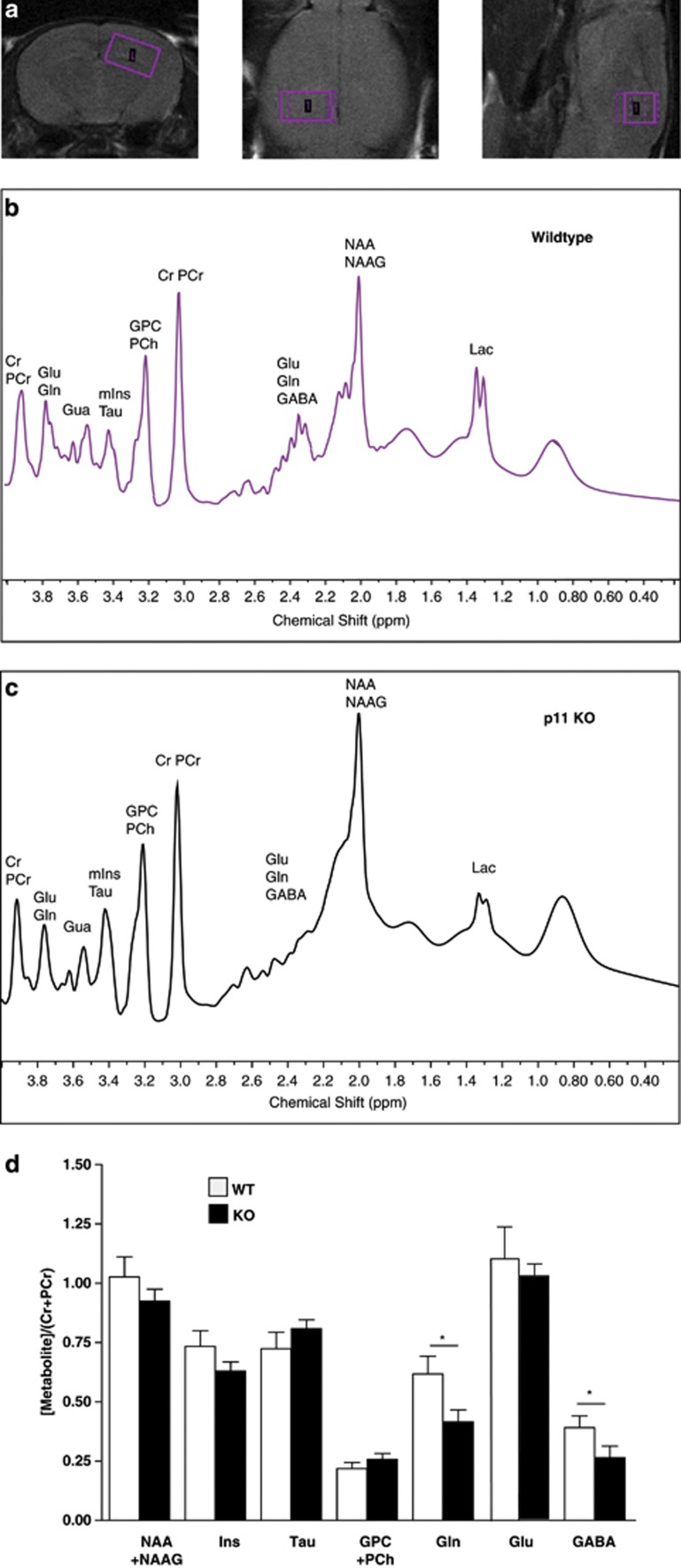
Figure 4.
Reduced hippocampal inhibitory transmitter detected by in vivo proton magnetic resonance spectroscopy (1H-MRS). Representative MRI (magnetic resonance image) featuring coronal, axial and sagital slices through a mouse brain (a). Placement of the voxel, the volume of interest (VOI) sized 3.0 × 1.8 × 1.8 mm3, for spectroscopy in the hippocampus indicated by the box. 1H-MR spectra acquired from the voxel centered in the hippocampus of WT (b) and p11KO mouse (c). Mean neurochemical concentration in WT (white bars) and p11KO mice (filled bars) (d). Relative concentrations of glutamine and GABA were reduced in the hippocampus of p11KO mice when compared to WT mice. Data are presented as means±s.e.m. for WT (n=7); P11KO (n=8). NAA+NAAG: N-Acetylaspartate+N-Acetylaspartatylglutamate, Ins: Inositol, Tau: Taurine, Glu: Glutamate, Gln: Glutamine, GABA; gamma-amino butyric acid, GPC+PCh: GlyceroPhosphocholine+Phosphocholine, WT: wild type mice, P11KO: p11 knock-out mice. *P<0.05.

