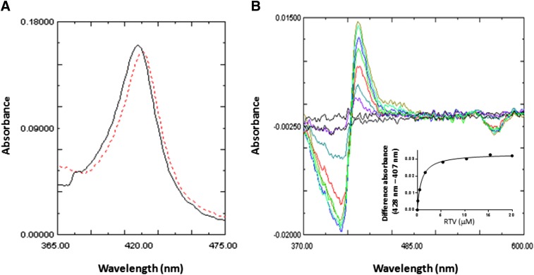
Fig. 6.
Spectra for RTV binding to CYP2B6. (A) Absolute spectra for CYP2B6 in the absence (solid line) or presence (dashed line) of 5 μM RTV. A shift of the maximal absorbance of 5 nm was observed. (B) RTV binding to CYP2B6 leading to a type II binding spectrum. The final concentrations of RTV added to the sample cuvette were 0.3, 0.9, 2.4, 5.4, 10.4, 15.4, and 20 μM. The inset shows the difference in the absorbance between the peak and the trough plotted versus the corresponding free RTV concentrations. The amount of RTV bound to 1 μM CYP2B6 at the lower concentrations was subtracted from the amount added, and the free concentrations of RTV used for the plot were 0.16, 0.5, 1.67, 5.4, 15.4, and 20 μM. The data points were fit to the Michaelis-Menten equation. The Ks was estimated to be 0.85 μM RTV.

