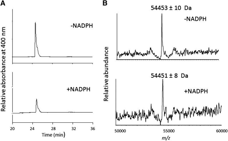
Fig. 7.
HPLC separation of the CYP2B6 prosthetic heme group after inactivation by RTV. (A) HPLC elution profiles monitored at 400 nm for the prosthetic heme in the absence or presence of NADPH. (B) The deconvoluted mass spectra of CYP2B6 incubated with RTV in the absence or presence of NADPH. The experimental procedures are described in the Materials and Methods.

