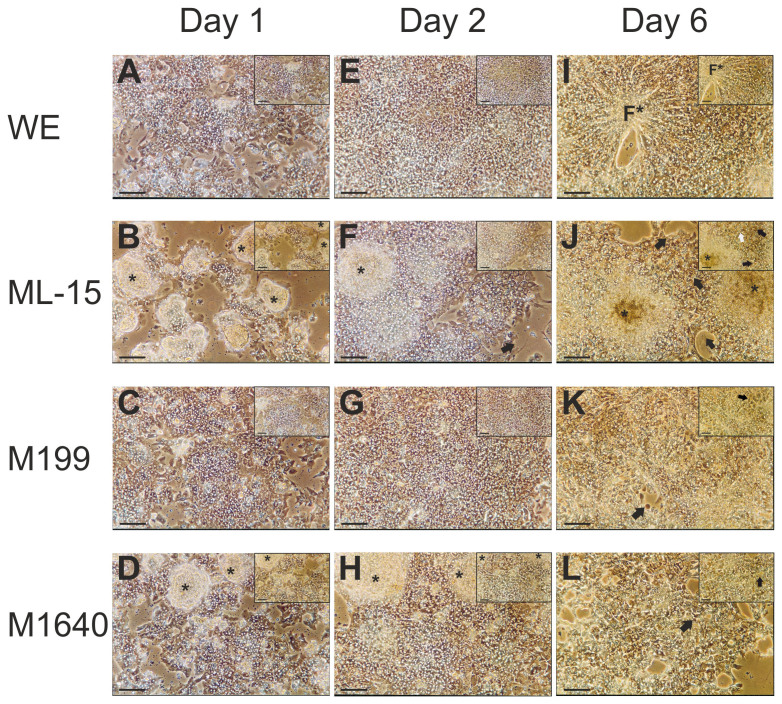
Figure 5. Phase contrast photomicrographs showing gross morphology of primary porcine hepatocytes cultured in four separate chemically-defined medium formulations.
Phase contrast micrographs showing gross morphology of primary porcine hepatocytes at day 1 (16 hours post-seeding), 2 and 6 in serum-free, chemically defined WE, ML-15, M199 or M1640 medium (pictures (A–L) ×100; scale bar 100 μm – inserts ×200; scale bar 50 μm). These images show the influence of test media on the attachment efficiency or degree of confluency on culture day 1. Cells grown in both WE (A, E, I) and M199 (C, G, K) media appear as almost confluent monolayers whereas ML-15 (B, F, J) and M1640 (D, H, L) maintained cells form partial monolayers with discrete colonies of piled up cells (denoted by asterisks). As expected, the presence of non-hepatocytes in areas of low cellular density is apparent, as is the presence of bile canaliculi by highly refractive cell borders under phase contrast (C, G). After 6 days, note the appearance in WE-culture of discrete foci (denoted by F*) of small aggregates with a radiating cord-like arrangement of hepatocytes, reminiscent of the in vivo cell microenvironment. However, both cell detachment and poor overall morphology compared with day 2 cultures was evident. The same was true to different degrees of degeneration for culture under the other tested media formulations as seen by cell detachment, general loss of delineating cell borders and the appearance of apoptosis, with cell blebbing (black arrows) and the appearance of lipid droplets and/or vacuoles (white arrows). Interestingly, cell colonies evident at earlier time-points under M1640 culture were no longer present.

