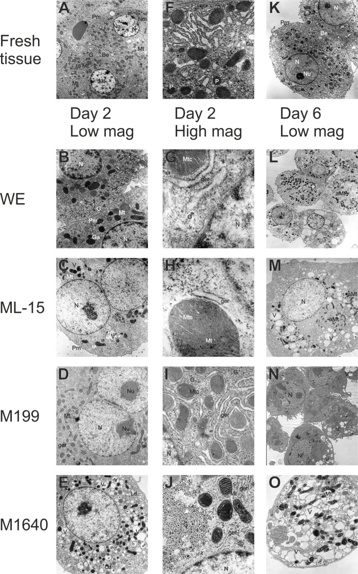
Figure 6. Transmission electron microscopy of fresh liver tissue and isolated hepatocytes.
Transmission electron micrographs of fresh liver tissue ((A, K) ×3800, (F) ×17000) and isolated hepatocytes exhibited common fine structural features. Round nuclei (N) with nucleoli (Nu) and well-defined plasma- (Pm), nuclear (Nm) membranes and lysosomes (L), abundant mitochondria (Mt) and intact mitochondrial cristae (Mtc) and granular endoplasmic reticulum (ger) interspersed between the mitochondria. Smooth endoplasmic reticulum and golgi apparatus (Ga), peroxisomes (P) are evident, as well as numerous dense glycogen (G) β-particles and rosettes (α-particles) between smooth endoplasmic reticulum and in close proximity to mitochondria. Bile canaliculus-like structures (Bc) are formed between adjacent hepatocytes in both fresh liver tissue and in isolated hepatocytes. Pictured hepatocytes correspond to phase contrast micrographs of day 2 and 6 displayed in Figure 5. The photomicrographs show areas of high morphological representation and were taken at the following magnifications appropriate for visualization: ×3800 (M, N), ×5000 (B, E, L), ×6500 (C, D), ×10000 (O), ×28000 (I, J) and ×75000 (G, H). Based on TEM pictures, cultures were subsequently assessed for the relative frequencies of cytoplasmic organelles and bile canaliculus-like structures on days 2 and 6 of culture for each test medium (as summarised in Table 1). Depending on the culture medium tested, a proportional reduction of intact cytoplasmic structures was observed in parallel with the occurrence of asymmetric mitochondrial swelling (aMt), the formation of lipid droplets (Ld) and extensive vacuolation (V).

