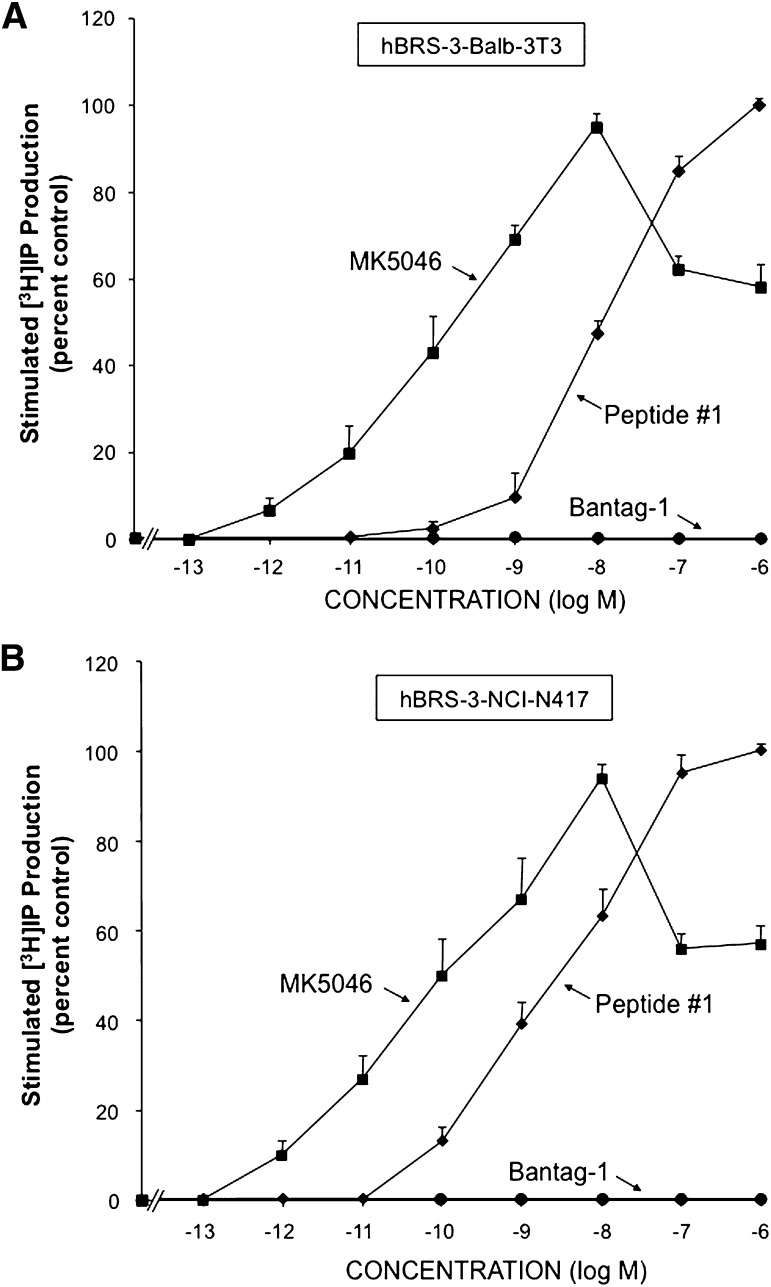
Fig. 3.
Ability of the agonists peptide #1 and MK-5046 or the putative antagonist Bantag-1 to stimulate [3H]IP generation in two different cell types containing hBRS-3: (A) hBRS-3 Balb 3T3 and (B) NCI-N417. After loading the cells with 3 μCi/ml myo-[2-3H]inositol (as described under Materials and Methods), each cell type was incubated with each peptide at the indicated concentration for 60 minutes at 37°C. The [3H]IP measurement was determined as described under Materials and Methods. The results are the mean ± S.E.M. from at least four experiments, and in each experiment the data points were determined in duplicate. The results are expressed as the percentage of stimulation caused by the maximal effective concentration of peptide #1 (1 μM). (A) With hBRS-3 Balb 3T3 cells, the maximal stimulated [3H]IP value by 1 μM peptide #1 was 14,695 ± 1481 dpm, and the control value was 4142 ± 407 dpm (n = 5). (B) With NCI-N417 cells, the maximal stimulation of a 1 μM concentration of peptide #1 was 2897 ± 440 dpm, and the control value was 1251 ± 199 dpm (n = 24).

